X-ray, or radiography, is the oldest and most common form of medical imaging. An X-ray machine produces a controlled beam of radiation, which is used to create an image of the inside of your body. This beam is directed at the area being examined. After passing through the body, the beam falls on a piece of film or a special plate where it casts a type of shadow. Different tissues in the body block or absorb the radiation differently. Dense tissue, such as bone, blocks most of the radiation and appears white on the film.
Soft tissue, such as muscle, blocks less radiation and appears darker on the film. Often multiple images are taken from different angles so a more complete view of the area is available. The images obtained during X-ray exams may be viewed on film or put through a process called digitizing so that they can be viewed on a computer screen.

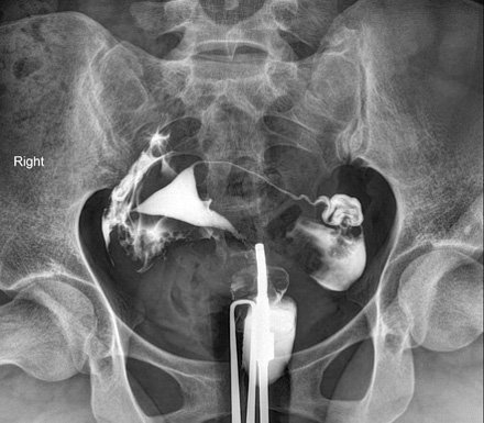
HSG X-ray

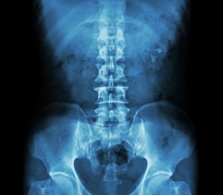
IVP X-ray

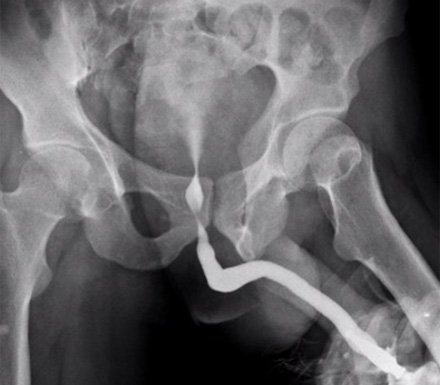
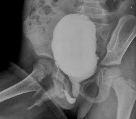
RGU X-ray

